Strictly at the Border
Why does Fischer's vascular deposit stop at the pia when modern angiopathy does not?
Fischer's sixth stage, the leptomeningeal compartment it never entered, and what a silver impregnation could not have shown
Abstract
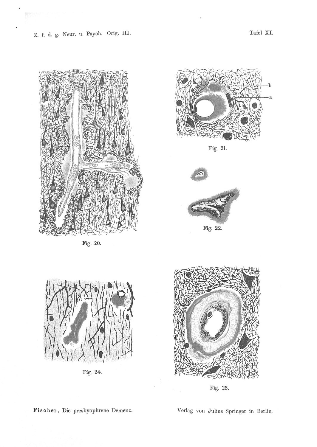
In 1910 Oskar Fischer described cortical vessels ensheathed by a dense radial deposit, called it Pelzbesatz — fur trim — and made it the sixth of his eight stages. Figures 21 and 23 of Tafel XI are, to a modern eye, unmistakable: a lumen, a wall, and a dense investment standing off it like a pelt. The stage is routinely and correctly identified as cerebral amyloid angiopathy, described in human cortex three-quarters of a century before the peptide was purified from it.
This paper is about a sentence that identification cannot accommodate.
"daß die Drusen streng mit dem Rande des Nervengewebes aufhören, daß auch diejenigen Rindengefäße, welche von den Drusen auf lange Strecken eingeschlossen werden, diese Umkleidung mit dem Moment verlieren, wie sie in die Meningen oder in die weiße Substanz eintreten."
("that the drusen stop strictly at the border of the nervous tissue; that even those cortical vessels which are enclosed by drusen over long stretches lose this investment the moment they enter the meninges or the white matter.")
Fischer's vascular deposit quits at the pia. Modern cerebral amyloid angiopathy does not. It is defined, in the standard classification, as a β-amyloidosis of the leptomeningeal and cortical vessels of the elderly, and its two sporadic types are separated by whether cortical capillaries are involved — both types show deposits in leptomeningeal vessels (Thal et al., 2002). In the severe disease that comes to neurosurgical attention, the burden is heaviest in medium- and large-calibre leptomeningeal vessels (Poyuran et al., 2019). The compartment Fischer's deposit refuses to enter is the compartment in which the modern lesion is worst.
Something must give. This paper sets out the three ways it can, tests each, and argues that the right answer is the one that is least flattering to the historical record and most useful to the modern reader.
The first possibility is that Fischer was wrong — that he under-sampled the meninges, which are stripped or torn in ordinary brain removal and are the first thing lost in a poorly handled autopsy. This is the null hypothesis and it cannot be excluded. It is weakened, though not defeated, by the specificity of his claim: he does not report an absence of findings in the meninges, he reports vessels followed out of the cortex and losing their investment at a boundary. That is a statement about continuity along a single vessel, and it is not the kind of statement that under-sampling produces.
The second is that his material differed — that the cortical and leptomeningeal compartments really did behave differently in a 1910 Prague autopsy population, for reasons of age structure, cause of death, or APOE allele frequency that cannot now be recovered. We cannot test it and we do not pursue it.
The third is that his stain was reading a different object, and this is what we argue. Bielschowsky silver impregnates neurofibrils. It does not stain amyloid, and Fischer knew what his stain was for: his own 1912 solvent series concluded that the thread material is proteinaceous rather than lipoid, and his figures throughout are figures of fibrils. A dense radial fibrillar investment around a cortical vessel requires fibrils to be there — which is to say, it requires neuropil. Cross the pia and the neuropil ends. On this reading, what Fischer scored as Stage VI was not the amyloid in the vessel wall but the fibrillar response of the surrounding tissue to it, and the response necessarily stops where the tissue does. The boundary in his sentence is not a boundary of the deposit. It is the boundary of the thing his method could see.
If that is right, three consequences follow, and they are the substance of the paper.
Stage VI is not CAA. It is the neuritic corona of a perivascular deposit — the vascular counterpart of the neuritic plaque, related to CAA as the neuritic plaque is related to the diffuse one. Fischer's own scoring supports this: he scored Pelzbesatz in cerebellar vessels too, and the cerebellum is a territory where his parenchymal deposits are sparse, Stage III, and — his own emphasis — never carry clubs. A vascular finding that appears where the neuritic response is otherwise absent is a difficulty for this reading and is treated as such in Chapter 14 rather than smoothed away.
The oldest morphological series bearing on vascular amyloid is therefore a series about something else, and the priority claim usually made for Fischer in this compartment should be narrowed accordingly. He did not stage CAA in 1910. He staged the tissue's reaction to it, which nobody stages now.
And that suggests a measurement the modern field does not make. Cerebral amyloid angiopathy is graded by the amount of amyloid in the wall (the Vonsattel scale and its descendants) and phased by which vessels are involved. Neither instrument scores the parenchymal response around the affected vessel. If Fischer's fur is the perivascular analogue of the neuritic corona, then a vessel-level dystrophy score is available, has never been constructed, and would stand to the vascular compartment as per-plaque dystrophy scoring stands to the parenchymal one — where it is known that local injury climbs across the clinical course while burden does not.
We close with a graded ledger of sixteen propositions, five falsifiers and four experiments in order. The first is a re-reading of Fischer's own plate against the modern question — whether the radial investment in Figs 21 and 23 is continuous with the neuropil, as a neuritic corona must be, or free-standing in the wall, as amyloid would be. That is answerable from a public-domain lithograph, and it decides the paper.
What this paper does not claim. It does not claim that Fischer failed to see vascular amyloid; the material in the wall was almost certainly there, and the modern identification of Stage VI with the vascular compartment is correct as far as compartment goes. It does not claim that his boundary sentence is more reliable than modern immunohistochemistry, which it plainly is not. And it does not claim to resolve the direction of the perivascular route, which is a live dispute this paper deliberately stays out of. The claim is narrower: that a well-known identification has been made between a modern lesion and a historical figure of a different object, that the discrepancy which reveals it has been in print since 1910, and that reading it correctly turns a priority footnote into an unmade measurement.
Part One — The Sentence
1. What Fischer Reported at the Vessel
Fischer's sixth stage is the vascular one, and he treats it as more than a morphological variant. It carries an argument.
The description is of cortical vessels invested by the same thread material that forms his drusen, arranged radially, standing off the wall. His word for it is Pelzbesatz, fur trim, and it appears in his case protocols as a scored finding — a thing he looked for and recorded case by case, including in cerebellar vessels. He also describes the deposit growing through the wall, which is what makes it a destruction rather than an ensheathment: "pelzartige Destruktion der Gefäßwand."
The argument he builds on it is directed against the idea that the drusen are ordinary catabolic residue:
"Ein Abbauprodukt, das so schön regelmäßig die Gefäße umscheidet und dabei die Wand des Gefäßes so zierlich und regelmäßig destruiert, würde etwas ganz Sonderbares darstellen."
("A breakdown product which so beautifully and regularly ensheathes the vessels, and in doing so destroys the vessel wall so daintily and regularly, would be something quite extraordinary.")
He is arguing from order. A waste product should not organise itself. The regularity of the vascular investment is, for him, evidence that the material is not debris — the same argument he makes from the orderly stage series in the parenchyma, transferred to the vessel.
And then he adds the boundary condition, which is the subject of this paper.
2. The Boundary Condition
"daß die Drusen streng mit dem Rande des Nervengewebes aufhören, daß auch diejenigen Rindengefäße, welche von den Drusen auf lange Strecken eingeschlossen werden, diese Umkleidung mit dem Moment verlieren, wie sie in die Meningen oder in die weiße Substanz eintreten."
Four features of the sentence are worth marking, because they determine how much weight it can carry.
It is a claim about continuity, not about prevalence. Fischer is not reporting that he searched the meninges and found nothing. He is reporting that he followed individual vessels — ones enclosed by drusen auf lange Strecken, over long stretches — and watched the investment stop. That is a within-vessel observation, and it is the kind of observation a sampling failure does not generate. A meninges that was torn off in removal produces silence, not a described transition.
It has two termini and they are of different kinds. The investment is lost at the meninges and at the white matter. The second is easy: white matter is not neuropil in the cortical sense, and Fischer separately records that the drusen do not occur in white matter at all. The first is the anomaly. But the fact that he gives both in one clause is informative: he is describing a single rule — the material stops where the grey stops — and the meningeal terminus is one instance of it rather than a separate finding about the meninges.
It is used as an argument, which means he thought it mattered. The sentence appears in his discussion of the perivascular route. He notes, correctly, that "es gehört zur Regel, daß Abbauprodukte den Weg der perivaskulären Lymphräume nehmen" — it is the rule that breakdown products take the way of the perivascular lymph spaces — and then uses the boundary to argue that his drusen are nevertheless in the tissue rather than in the perivascular drainage, because material travelling in a perivascular space would not stop at the pia. His inference is wrong, on the modern account, but the observation he draws it from is stated precisely because it was load-bearing for him.
It is unhedged. Fischer hedges when he is inferring — the ich betone hier das „könnte" of his colour argument is the model — and states flatly when he is reporting. This is stated flatly.
3. What Modern CAA Actually Is
The comparison requires the modern lesion to be described in its own terms rather than in a summary.
Cerebral amyloid angiopathy is a β-amyloidosis affecting leptomeningeal and cortical vessels in the elderly. In a sample of 41 CAA cases including 16 with Alzheimer's disease, together with 28 controls, two sporadic types are distinguishable and the distinction is not one of severity. Type 1 shows immunohistochemically detectable Aβ in cortical capillaries as well as in leptomeningeal and cortical arteries, arterioles, veins and venules. Type 2 shows the same distribution with the exception of cortical capillaries. The ratio of the two types does not shift with the severity of Alzheimer β-amyloidosis, with degree of CAA severity, or with age, which is the argument that Type 1 is not simply a late Type 2: they are different entities. And the genotype separates them — the APOE ε4 allele frequency is more than four times greater in Type 1 than in Type 2 or controls, while Type 2 carries a higher ε2 frequency (Thal et al., 2002).
Two things in that description matter here.
Leptomeningeal involvement is definitional and is common to both types. It is not a late feature, not a variant, and not restricted to severe cases. It is in the first clause of the definition.
The compartment that separates the types is the cortical capillary — the smallest, deepest, most parenchymal vessel in the set. The classification's own axis of variation runs in the opposite direction from Fischer's boundary: it distinguishes cases by how far in the deposit goes, while Fischer's sentence is about how far out it does not.
Severity confirms the same geography. In biopsy- and resection-confirmed CAA presenting as intracerebral haemorrhage, moderate-to-severe vascular amyloid showed predominant involvement of medium (200–500 µm) to large (>500 µm) leptomeningeal vessels, with fibrinoid necrosis in four of seven cases; amyloid in small-to-medium intracortical vessels produced parenchymal microhaemorrhages instead, and it was vessel calibre rather than amyloid grade that determined the size of the bleed (Poyuran et al., 2019).
The clinical weight of CAA — the haemorrhages, the reason the lesion is graded at all — sits in the compartment Fischer's deposit does not enter.
4. The Three Ways Out
Only three readings are available, and it is worth listing them before arguing for one, because two of them are boring and the paper must not be allowed to skip them.
(a) Fischer under-sampled the meninges. The leptomeninges are the most easily lost tissue in a brain autopsy; they strip with the dura, they tear on removal, and they are not reliably present on a cortical block cut for parenchymal study. If they were largely absent from his sections, he would find no vascular investment beyond the cortical surface for the trivial reason that he had nothing to look at there.
(b) His population differed. Allele frequencies, age structure and cause of death in a Prague asylum population of 1905–1910 are not those of a modern autopsy series, and CAA type is genotype-linked. This is unfalsifiable now and is not pursued beyond noting it.
(c) His stain was reading a different object. Bielschowsky silver demonstrates neurofibrils. If the radial investment he drew is a fibrillar structure — the neuropil's response to a perivascular deposit rather than the deposit itself — then it must end where the neuropil ends, and the pial boundary is a property of the observer's method rather than of the disease.
Chapters 5 to 9 test (a) and argue (c). The test that separates them is available in a public-domain lithograph and is set out in Chapter 15 as the first experiment.
Part Two — Testing the Readings
5. Against Under-Sampling
The under-sampling hypothesis is the null and it deserves the strongest form.
Fischer's material was cortex. His question was the cortical deposit; his stain was chosen for cortical fibrils; his 275 brains were sampled for a study of the cortical grey. Meninges adherent to a cortical block are a nuisance in that work, and a technician trimming blocks for silver impregnation has every reason to remove them. If the leptomeningeal compartment was simply not on the slide, the boundary sentence is an artefact of trimming.
Three considerations weigh against it, none decisive alone.
The sentence describes a transition, not an absence. This is the strongest point and it has been made already: Fischer reports vessels enclosed over long stretches that lose their investment at the moment of entry into the meninges. To write that sentence he had to have material on both sides of the boundary in the same section. A trimmed-away meninges gives you no transition to describe.
He reports the same rule at the white-matter boundary, where trimming is not a plausible explanation — nobody trims the white matter off a cortical block — and where he independently established that drusen do not occur at all. One rule with two termini, one of which is verifiable within his own dataset, is more likely to be a real rule than a coincidence of two artefacts.
He scored vascular involvement as a protocol item across cases, including in the cerebellum. A finding recorded case by case is one he was actively looking for, which raises the prior that he would have noticed and remarked on meningeal vessels if his sections carried them and they were invested.
What would settle it. Nothing in the printed record can, because the sections are gone and the question is about what was on them. The plate is the only surviving evidence, and Chapter 15's first experiment asks the plate a different question — not "were the meninges present?" but "is the investment continuous with the neuropil?" — precisely because the sampling question is unanswerable and the object question is not.
The under-sampling reading is therefore entered in the ledger as not excluded and is carried through the rest of the paper as a live alternative. A reader who prefers it should note that it leaves the modern identification of Stage VI with CAA intact and makes this paper unnecessary, which is a legitimate position and is stated as such in Chapter 17.
6. What Bielschowsky Silver Demonstrates
The case for the third reading begins with the method, and the method is not in dispute.
Bielschowsky's silver impregnation is a neurofibrillar method. It is the technique on which the entire 1907–1912 corpus rests, it is what allowed Fischer to draw the thread structure of the drusen at all, and it is what allowed him to trace club-shaped axonal swellings to their parent fibres in thick sections. It is a stain for the fibrous cytoskeletal architecture of nervous tissue.
It is not an amyloid stain. Congo red was not applied to brain tissue for this purpose until decades later; thioflavin later still; immunohistochemistry later again. Nothing in Fischer's panel — haematoxylin-eosin, van Gieson, Weigert's myelin and neuroglia methods, polychrome and tannin methylene blue, Marchi, Cajal, Levaditi — is a specific amyloid method either.
This is not a criticism of Fischer. He knew what he had and he was explicit that the threads themselves were the object: his 1912 solvent series, run against Marinesco's claim that the substance was lipoid, found the threads dissolved by neither alkalis, acids nor fat solvents, and concluded "es kann sich demnach kaum um etwas anderes als eine eiweißartige Substanz handeln" — it can accordingly hardly be anything other than an albuminous substance. He was studying a protein-fibrillar object with a protein-fibrillar method, correctly.
The consequence for the vascular stage is simply stated. Whatever Fischer's Pelzbesatz is, it is something a neurofibrillar impregnation can render as a dense radial array. That is a strong constraint, and it is a constraint the amyloid in a vessel wall does not obviously satisfy.
7. The Object That Requires Neuropil
Put the constraint together with the boundary and the reading assembles itself.
A radial fibrillar array around a cortical vessel, visible in a neurofibrillar stain, terminating exactly at the border of the nervous tissue, is what you would expect if the array is made of the tissue's own fibres. Fibrils are contributed by neurites; neurites belong to the neuropil; the neuropil stops at the pia and does not exist in white matter in the cortical sense. Every term of Fischer's rule is satisfied by an object of that kind, and the rule is satisfied automatically rather than as a further fact requiring explanation.
The modern name for the parenchymal version of this object is the neuritic corona: the ring of dystrophic neurites that surrounds a dense-core plaque and gives the neuritic plaque its name. Fischer described that too, at length, and it is the basis of his club-neurite rule. The proposal here is that his Stage VI is the same relationship transposed to a vessel — a perivascular deposit with a neuritic response around it, of which his stain shows the response.
There is a nice consistency check available inside his own data. Fischer's central quantitative claim in the parenchyma is that the club-shaped swellings cluster at the middle stages and are absent at both extremes of the series. If the vascular fur is a neuritic response, it belongs to the same family of phenomena as the clubs, and it should be subject to the same conditionality — present where there are neurites in a condition to respond, absent where there are not. That is a prediction, it is stated in Chapter 14, and it has a specific vulnerability described there.
8. Where the Peptide Actually Came From
An objection has to be met here, because it looks fatal and is not.
The peptide that defines the modern disease was purified from the vascular compartment first. Glenner and Wong obtained it in 1984 from cerebrovascular deposits in meningeal vessels; Masters and colleagues obtained it from plaque cores the following year, and the identity of the two established the continuity between the compartments (Glenner & Wong, 1984; Masters et al., 1985).
Meningeal vessels. The compartment Fischer's deposit does not enter is the compartment from which the founding preparation was made — and it was chosen precisely because meningeal vessels can be stripped and cleaned, giving a preparation far less contaminated by parenchyma than any cortical sample.
This does not contradict the reading; it sharpens it. The 1984 preparation isolated amyloid, from a compartment where amyloid is abundant and where neuropil is absent. Fischer's preparation rendered fibrils, in a compartment where fibrils are abundant. The two methods, applied to the same disease, pick out different objects with different distributions, and the distributions differ in exactly the way the two methods predict. The founding purification is therefore evidence for the claim that the leptomeningeal compartment carries vascular amyloid in quantity — which is what makes Fischer's silence there require explanation in the first place.
9. Fischer's Own Inference, and Why It Fails
For completeness: Fischer used the boundary to argue that his drusen were not travelling in the perivascular drainage. The argument runs that catabolic products characteristically take the perivascular route, that material in that route would not respect the tissue boundary, and that his material does respect it, so his material is in the tissue.
On the modern account the perivascular route is real and central — amyloid-β drains from the interstitium along vascular basement membranes, and deposits in the wall when that drainage fails — so his conclusion is wrong. But notice the structure of his error. He inferred a fact about the deposit from a boundary that belonged to his stain. That is the same class of error this paper attributes to the modern reading of his sixth stage, running in the opposite direction: he read a methodological boundary as biology, and the century that followed read his methodological object as the biological lesion.
Both errors have one source. Nobody asked what the silver was showing.
Part Three — Consequences
10. What Stage VI Is, If This Is Right
The proposal, stated as flatly as it can be:
Fischer's sixth stage is the perivascular neuritic corona, not cerebral amyloid angiopathy. It stands to CAA as the neuritic plaque stands to the diffuse deposit — the same underlying material, scored through the tissue's reaction to it rather than directly.
Three clarifications keep this from being either a bigger or a smaller claim than intended.
The compartment attribution is not disputed. Stage VI is vascular. Fischer was looking at deposits on and in cortical vessel walls, and vascular amyloid was almost certainly present in that material. What is disputed is which object the figure is a figure of.
This is not a demotion of Fischer. A century of readers has credited him with the earlier and easier finding — he saw the vascular amyloid — while overlooking the harder and more useful one, that the parenchyma mounts a graded, spatially organised response to a vascular deposit and that the response can be drawn. The second is worth more.
It is compatible with his material containing both. A cortical vessel bearing wall amyloid and a neuritic corona would give, in Bielschowsky silver, exactly the picture in Figs 21 and 23, with the amyloid contributing little to the image. The claim is about what the stain renders, not about what was in the tissue.
11. The Priority Claim, Narrowed
The standard sentence about Fischer and the vascular compartment is that he described cerebral amyloid angiopathy in 1910, decades before it was named and three-quarters of a century before the peptide was purified from it. On the reading argued here, that sentence should be narrowed.
What he described in 1910 was a vascular lesion in the cortical compartment, rendered through a neurofibrillar method, obeying a boundary rule that CAA does not obey. Calling it CAA imports the modern lesion's distribution — leptomeningeal, calibre-dependent, haemorrhage-producing — into a figure that shows none of those things and explicitly contradicts the first.
The narrowing costs Fischer a priority claim he is often given and hands him a different one that nobody has made: the first description of a graded parenchymal reaction to a vascular amyloid deposit. That is a real finding, it is in his figures, and — as Chapter 12 argues — it has no modern instrument.
There is a general lesson here that the historical literature keeps having to relearn. A historical figure is not a photograph of a disease; it is a photograph of what a method rendered. Identifying a modern lesion with a historical image requires knowing what the method could and could not show, and the identification of Stage VI with CAA has been made for a century without that step being taken.
12. The Measurement Nobody Makes
Cerebral amyloid angiopathy is assessed in two ways and neither of them looks at the tissue around the vessel.
Severity is graded by the amount and distribution of amyloid within the wall — the Vonsattel scale and its descendants, on which the field's principal correlations rest, including the APOE dose relationship: one ε4 allele raises the odds of moderate or severe CAA 2.9-fold and two copies 13.1-fold, independently of the presence of Alzheimer's disease (Greenberg et al., 1995).
Type and topography are assessed by which vessels carry deposit — capillary involvement separating Type 1 from Type 2, with the genotype associations noted above (Thal et al., 2002) — and by vessel calibre, which determines the clinical consequence more than the amyloid grade does (Poyuran et al., 2019).
Both instruments score the vessel. Neither scores what is happening to the neuropil around it.
That gap is conspicuous next to the parenchymal compartment, where scoring the tissue's response has been unusually informative. In dense-core plaques from temporal neocortex across forty subjects with symptom durations of four to twenty years, plaque burden was essentially stationary while per-plaque features climbed: SMI312-positive dystrophic neurites rose with duration at Kendall τ = 0.34, GFAP-positive astrocytes at τ = 0.30, and CD68-positive microglial activation at τ = 0.48, while IBA1-positive microglial number did not move at all (τ = 0.045) (Serrano-Pozo et al., 2016). The lesson of that result is that the count is the wrong variable and the local reaction is the right one.
A vessel-level dystrophy score is therefore available and unbuilt. Its terms would be: the fraction of the vessel's perimeter carrying dystrophic neurites, the density of those neurites per unit length of vessel, and the ratio of that density to the amyloid grade of the same segment. It would be scored on the same sections already cut for CAA grading, using antibodies already in routine use, and it would answer a question the existing instruments cannot: whether two vessels with the same wall burden are doing the same amount of damage to the tissue they run through.
Fischer scored the numerator of that ratio in 1910, case by case, and called it fur.
13. Why It Might Matter Clinically
One paragraph, kept short because it is the most speculative claim in the paper and the ledger grades it accordingly.
The vascular compartment is where the field's principal therapeutic strategy meets its dose limit. Amyloid-related imaging abnormalities arise from the vascular compartment and track CAA burden and APOE ε4. The instrument used to anticipate that risk is a measure of how much amyloid is in the wall. If the tissue's response to a given wall burden varies between individuals in the way that the parenchymal response demonstrably does, then wall burden is an incomplete predictor by construction, and a response measure would add information. This is a hypothesis about a possible clinical use, not a finding, and it is entered as beyond the evidence.
14. Where This Reading Is Weak
The paper's own difficulties, stated before the ledger rather than after it.
The cerebellar scoring is a problem. Fischer scored Pelzbesatz in cerebellar vessels. The cerebellum is a territory where his parenchymal deposits are sparse, assigned to Stage III, and — his emphasis — never carry clubs. If the vascular fur is a neuritic response, it should be scarce or absent exactly where the neuritic response is otherwise absent. Finding it there is a difficulty, and there are only two ways out, neither comfortable: either the vascular corona is generated by a different neurite population from the plaque-associated clubs, or the cerebellar vascular finding is a different object again. This is the strongest single objection to the paper and it is entered in the ledger as contested.
The transition sentence may be over-read. The whole case against under-sampling rests on Fischer describing a within-vessel transition. If that sentence is a generalisation dressed as an observation — a summary of "we do not see this in meninges" written in vivid form — the argument loses its best evidence. Nothing in the printed text settles this.
No modern study has looked. There is, as far as we can establish, no published quantification of perivascular dystrophic neurites stratified by wall amyloid grade in human cortex. The paper's central positive proposal therefore rests on an analogy with the parenchymal compartment rather than on a measurement.
The reading does not touch the direction dispute. Whether amyloid reaches the wall by failed perivascular drainage from the interstitium or by another route is live, and this paper's argument is neutral on it. Readers looking for an answer there will not find one, and the neutrality is deliberate: a paper about what a stain could see should not adjudicate a question about flow.
Part Four — The Ledger
15. A Graded Ledger
Established — directly evidenced in human material or by convergent human and animal data. Supported — good evidence with a stated dependency. Inference — consistent with the evidence, not directly demonstrated. Contested and beyond the evidence are used where they are more honest than a grade.
Three rows deserve comment.
Row 7 is the finding, and it is not a proposal. That Fischer's boundary and the modern definition of CAA are incompatible is a matter of reading two texts. Everything after row 7 is about why, and the paper's preferred answer is graded as an inference in row 11 rather than as a result.
Row 8 leaves the paper unnecessary if it is true. A reader who concludes that the meninges were simply not on the slide should stop at row 8, keep the conventional identification of Stage VI with CAA, and disregard Parts Three and Four. That position is honest and the paper cannot refute it from printed material. What it can do is offer a test that distinguishes the two — Experiment 1 — and note that the sampling reading has to explain away the transition described in row 3.
Row 12 is the objection this paper does not answer. Cerebellar Pelzbesatz in a territory with no clubs is exactly what the neuritic-corona reading should not predict. It is entered as contested, it is discussed in Chapter 14, and it is the first thing a critic should press.
Each claim below carries a tier and, where it is not settled, the observation that would settle it. 9 claims · 2 not yet settled
Strong — Modern CAA involves leptomeningeal vessels by definition, in both sporadic types
Thal 2002, n = 41 CAA cases + 28 controls
What would settle it. — not in dispute
Strong — Severe CAA is heaviest in medium-to-large leptomeningeal vessels
Poyuran 2019, biopsy/resection-confirmed
What would settle it. —
Strong — APOE ε4 raises CAA severity in a dose-dependent way
Greenberg 1995, OR 2.9 for one copy, 13.1 for two
What would settle it. —
Strong — Bielschowsky silver is a neurofibrillar method and not an amyloid method
The method; Fischer's own use of it throughout
What would settle it. —
Strong — The founding purification of the peptide was made from meningeal vessels
Glenner & Wong 1984; Masters 1985
What would settle it. —
Strong — Scoring the parenchymal reaction is more informative than scoring the burden, in the parenchymal compartment
Serrano-Pozo 2016, n = 40; burden flat, per-plaque features climb
What would settle it. —
Established (supported, textual) — The statement describes a within-vessel transition, not an absence of findings
The wording — vessels enclosed auf lange Strecken losing the investment on entry
What would settle it. Evidence that the sentence is a generalisation rather than an observation
Contested — Fischer scored Pelzbesatz in cerebellar vessels, where his parenchymal deposits never carry clubs
1910
What would settle it. This is the strongest objection to row 11 and is unresolved
Weak (beyond the evidence) — A vessel-level dystrophy score would add information over wall amyloid grade
15, by analogy
What would settle it. Experiment 3
16. Predictions and Falsifiers
P1. In Figs 21 and 23 of Tafel XI, the radial investment will show continuity between the peri-vessel array and the surrounding neuropil, rather than a free-standing annulus separated from the tissue. Falsified by: a free-standing annulus with a clear zone between it and the neuropil, which is what a wall deposit rendered directly should look like.
P2. In modern human cortex, a silver impregnation applied alongside an amyloid immunostain on affected vessels will render the perivascular neuritic material and not the wall deposit. Falsified by: silver rendering the wall deposit as a radial array.
P3. Perivascular dystrophic neurite density will vary between individuals at matched wall amyloid grade — that is, wall grade will not determine the local reaction. Falsified by: a tight, near-deterministic relation between wall grade and perivascular dystrophy.
P4. Perivascular dystrophic neurite density will rise with symptom duration while CAA grade does not, mirroring the parenchymal dissociation. Falsified by: both rising together, or neither moving.
P5. In the cerebellum, vessels showing Pelzbesatz-like investment will lack the neuritic markers found around cortical vessels. Falsified by: equivalent perivascular neuritic dystrophy in cerebellar vessels — which would rescue Fischer's cerebellar scoring at the cost of row 11's coherence, and is the outcome this paper's critic should hope for.
17. Four Experiments, in Order
1. Re-read Tafel XI for continuity. The plate is public domain and the question is answerable from the lithograph: is the radial material continuous with the neuropil, or free-standing? Score Figs 20–23 blind to the hypothesis, with the parenchymal plates as internal comparators, since the neuritic corona of a dense-core plaque appears elsewhere in the same series and provides the positive control. This decides row 11 as far as historical material can, and it costs a week.
2. Silver and amyloid on the same vessel. Apply a Bielschowsky or modified silver impregnation and an Aβ immunostain to serial or double-labelled sections of CAA-affected cortex, and establish directly what the silver renders. Tests P2. This is a methods experiment on ordinary material and is the single most decisive item in the list; it is second only because the first is free.
3. Build the vessel-level dystrophy score. On sections already cut for CAA grading, quantify perivascular dystrophic neurites per unit vessel length and as a fraction of vessel perimeter, stratified by wall amyloid grade, calibre and compartment. Tests P3 and, with clinical durations attached, P4. This is the experiment that would turn the historical observation into a modern instrument.
4. The cerebellar question. Score cerebellar vessels for perivascular neuritic dystrophy alongside cortical vessels in the same brains. Tests P5 and addresses row 12 directly. Placed last because it is the one most likely to embarrass the paper, which is not a reason to omit it but is a reason to have the other three in hand first.
18. Limitations
The central proposal is an inference about what a lost preparation showed. No experiment can recover Fischer's sections. Experiment 1 interrogates a lithograph; Experiment 2 establishes what the method does in general, not what it did in his hands.
The under-sampling alternative cannot be excluded and is graded accordingly. If it is right, this paper is a long footnote.
The cerebellar scoring is unexplained. Row 12, Chapter 14, Experiment 4.
The vessel-level dystrophy score is proposed and not piloted. No feasibility work has been done, and post-mortem lability of the phospho-epitopes used for neuritic markers is a known obstacle in this class of measurement.
Nothing here bears on the direction of the perivascular route, and readers should not take the paper's use of the drainage literature as support for either side of that dispute.
19. Conclusion — The Boundary Was the Method's
Fischer followed cortical vessels invested in a dense radial fur, watched the fur stop at the pia, and wrote the observation down as a fact about the deposit. It was not. It was a fact about what a neurofibrillar impregnation can render, and the material his stain was rendering ends where the nervous tissue ends.
A century of readers has taken his figure for a picture of cerebral amyloid angiopathy, in a lesion whose modern definition begins with the leptomeningeal compartment his deposit never enters. The discrepancy has been in print, unremarked, since 1910. It is not a small one, and reading it correctly does two things.
It narrows a priority claim: he did not stage the vascular amyloid. And it hands over a different one, which is worth more, because nobody has taken it up. He staged the reaction — the graded, spatially organised response of the neuropil to a vascular deposit, scored vessel by vessel, case by case, in human tissue, and called Pelzbesatz because that is what it looks like.
The modern instruments for cerebral amyloid angiopathy score the wall. In the parenchymal compartment, the equivalent move — scoring the burden rather than the reaction — was shown a decade ago to be measuring the wrong thing: the count stands still across two decades of clinical course while the local injury climbs. There is no reason to expect the vessel to be different, and there is at present no way to find out, because the measurement Fischer made by eye in 1910 has no modern form.
Sources and Reproductions
Tafel XI of the 1910 monograph (published by Julius Springer, Berlin) is in the public domain and is reproduced from the original printing.
All German quotations are taken from the original papers and located by journal page in the text. Translations are the author's, with the German given alongside wherever the reading turns on it.
References
Glenner GG, Wong CW. Alzheimer's disease: initial report of the purification and characterization of a novel cerebrovascular amyloid protein. Biochemical and Biophysical Research Communications. 1984;120(3):885–890. DOI 10.1016/S0006-291X(84)80190-4
Greenberg SM, Rebeck GW, Vonsattel JP, Gomez-Isla T, Hyman BT. Apolipoprotein E ε4 and cerebral hemorrhage associated with amyloid angiopathy. Annals of Neurology. 1995;38(2):254–259. PMID 7654074. DOI 10.1002/ana.410380219
Fischer O. Die presbyophrene Demenz, deren anatomische Grundlage und klinische Abgrenzung. Zeitschrift für die gesamte Neurologie und Psychiatrie. 1910;3:371–471. (Mit 13 Tafeln.) Find this paper
Fischer O. Ein weiterer Beitrag zur Klinik und Pathologie der presbyophrenen Demenz. Zeitschrift für die gesamte Neurologie und Psychiatrie. 1912;12:99–135. (Mit 1 Tafel.) Find this paper
Masters CL, Simms G, Weinman NA, Multhaup G, McDonald BL, Beyreuther K. Amyloid plaque core protein in Alzheimer disease and Down syndrome. Proceedings of the National Academy of Sciences USA. 1985;82(12):4245–4249. DOI 10.1073/pnas.82.12.4245
Poyuran R, Mahadevan A, Arimappamagan A, Nandeesh BN, Nagappa M, Saini J, Narasinga Rao KVL, Chickabasaviah YT. Cerebrovascular pathology in cerebral amyloid angiopathy presenting as intracerebral haemorrhage. Virchows Archiv. 2019;474(2):235–245. PMID 30617647. DOI 10.1007/s00428-018-2505-z
Serrano-Pozo A, Betensky RA, Frosch MP, Hyman BT. Plaque-associated local toxicity increases over the clinical course of Alzheimer disease. The American Journal of Pathology. 2016;186(2):375–384. PMID 26687817. DOI 10.1016/j.ajpath.2015.10.010
Thal DR, Ghebremedhin E, Rüb U, Yamaguchi H, Del Tredici K, Braak H. Two types of sporadic cerebral amyloid angiopathy. Journal of Neuropathology and Experimental Neurology. 2002;61(3):282–293. PMID 11895043. DOI 10.1093/jnen/61.3.282
Genes named on this page: ApoE, apolipoprotein E; GFAP; CD68; AIF1 (Iba1), IBA1.